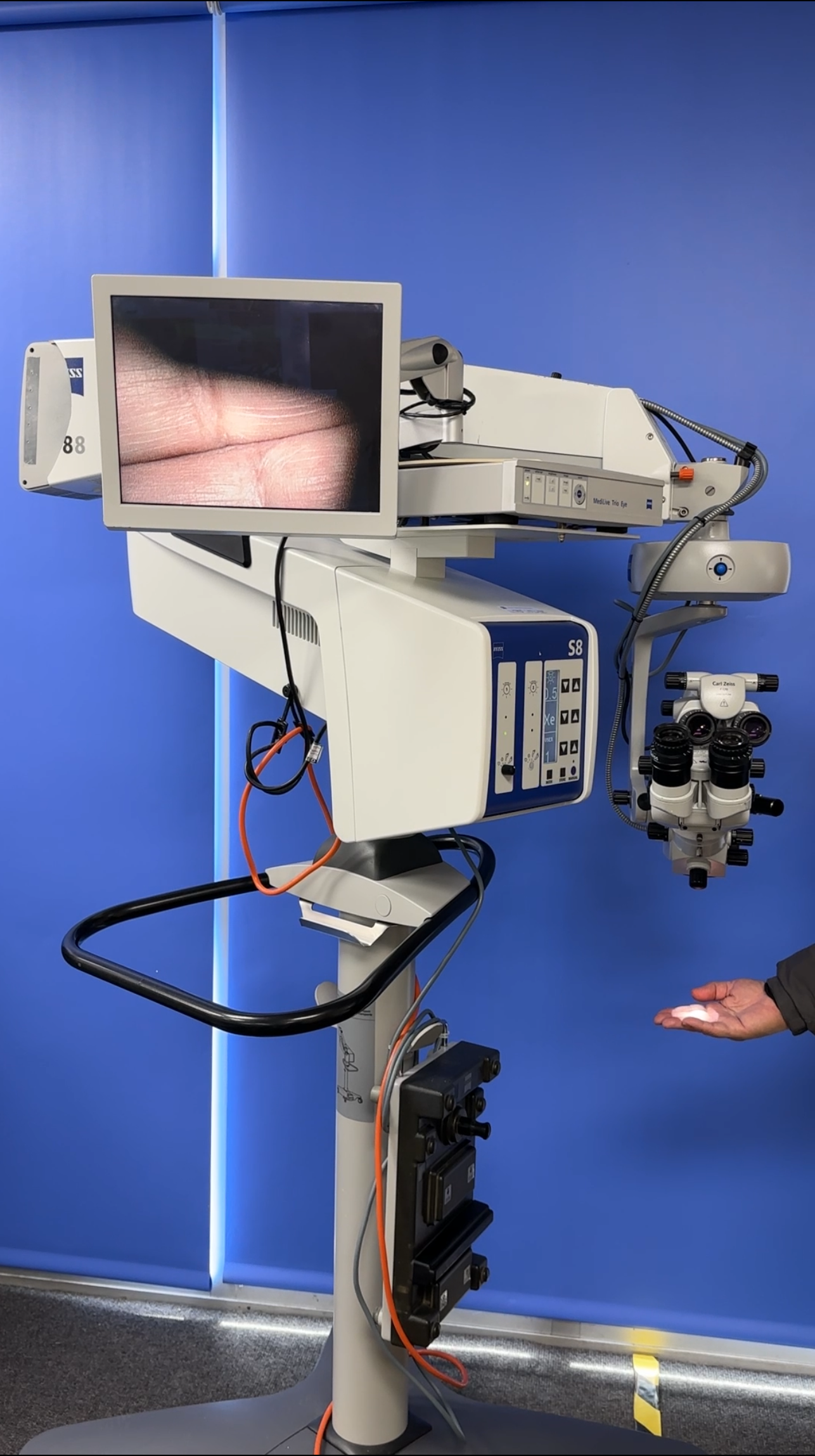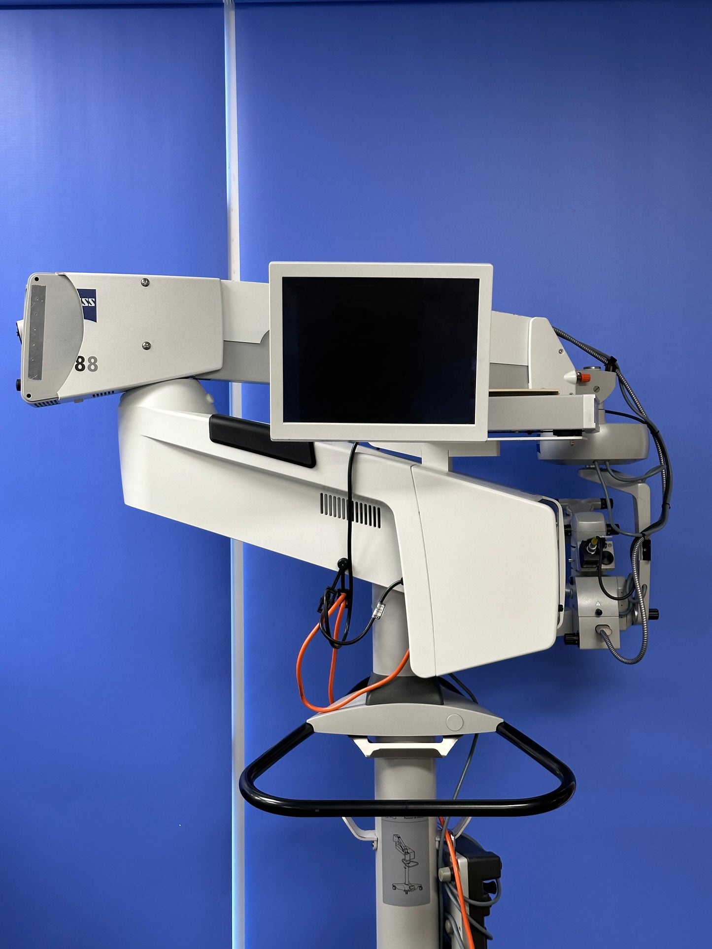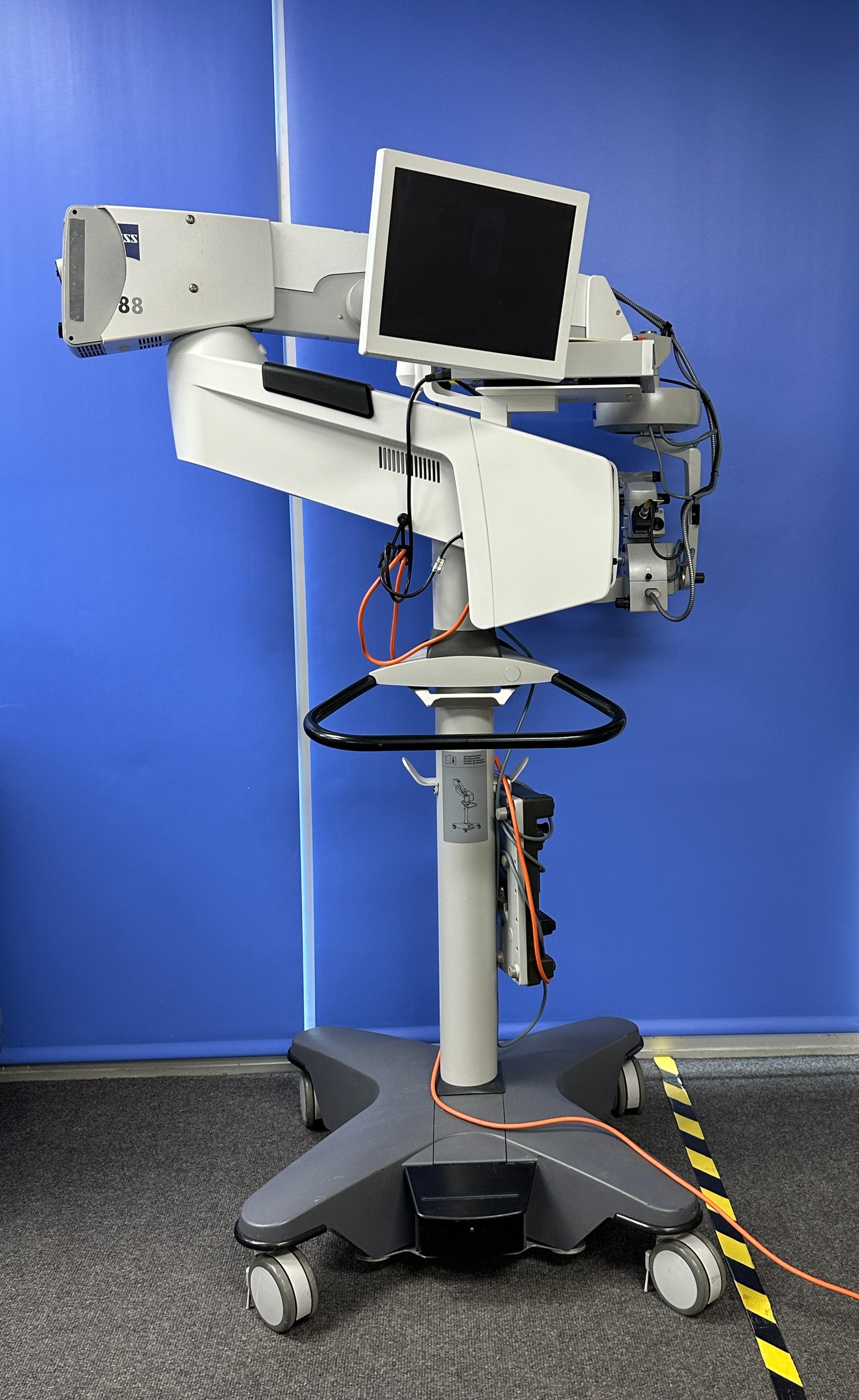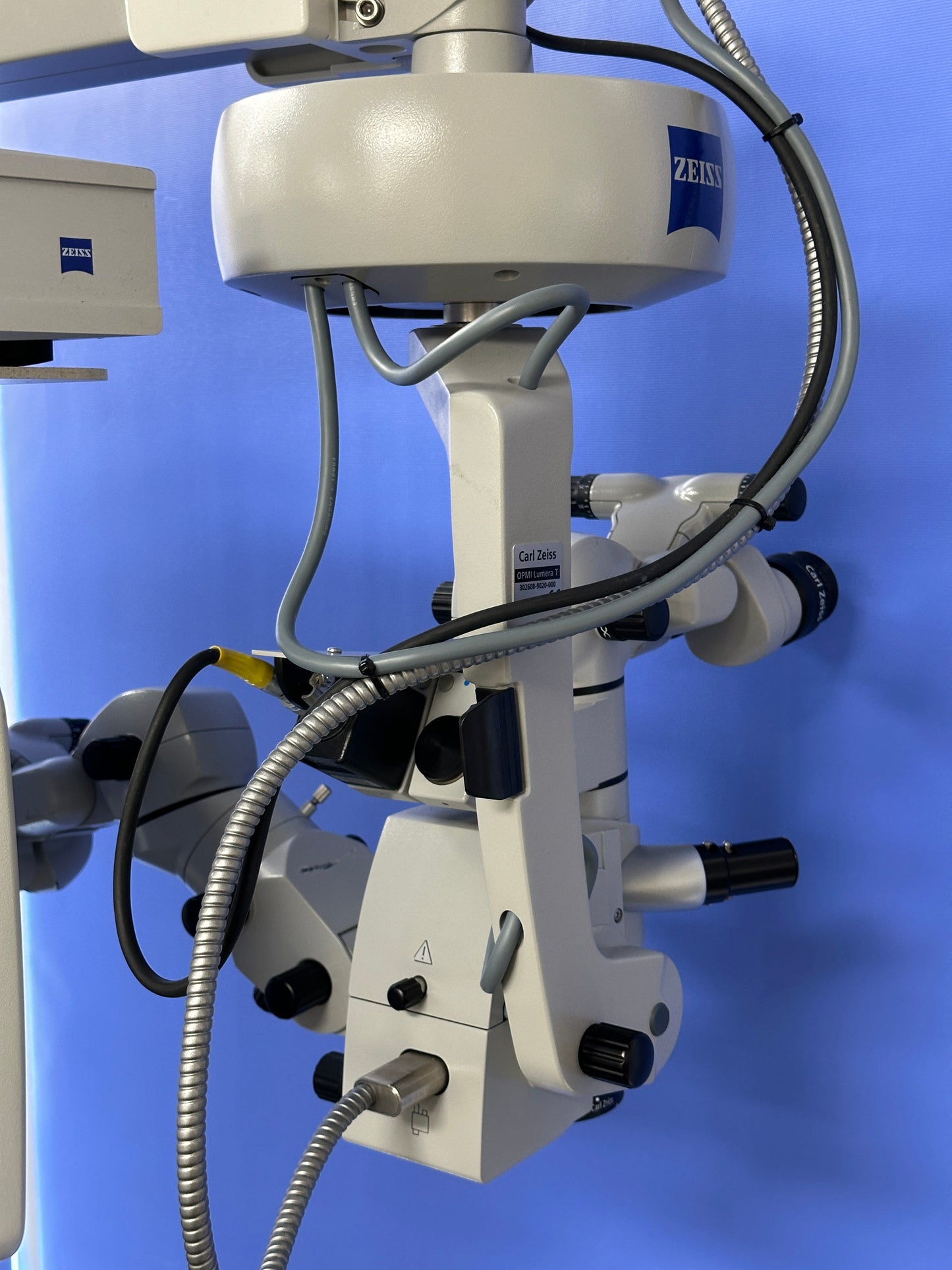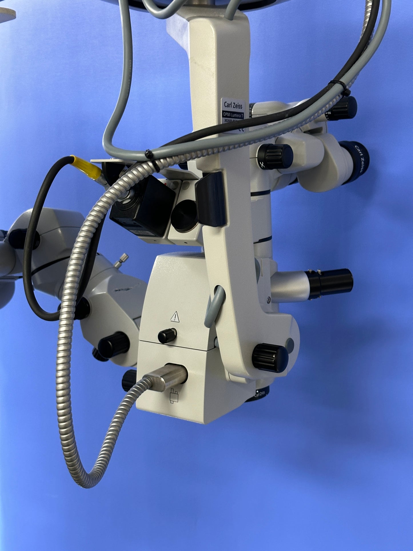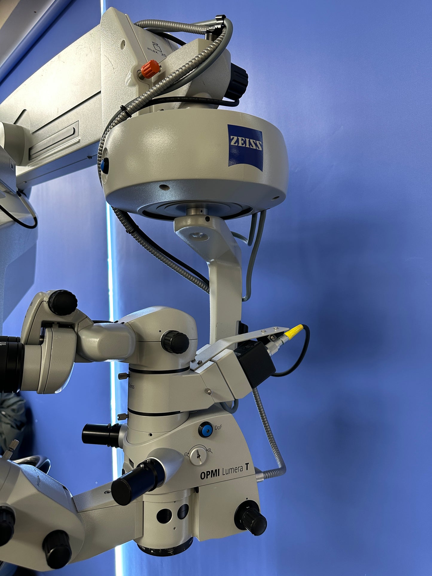


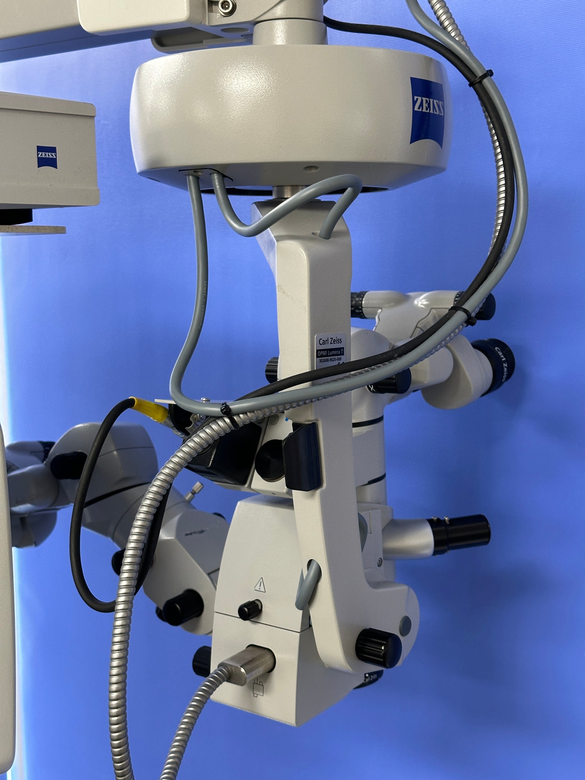
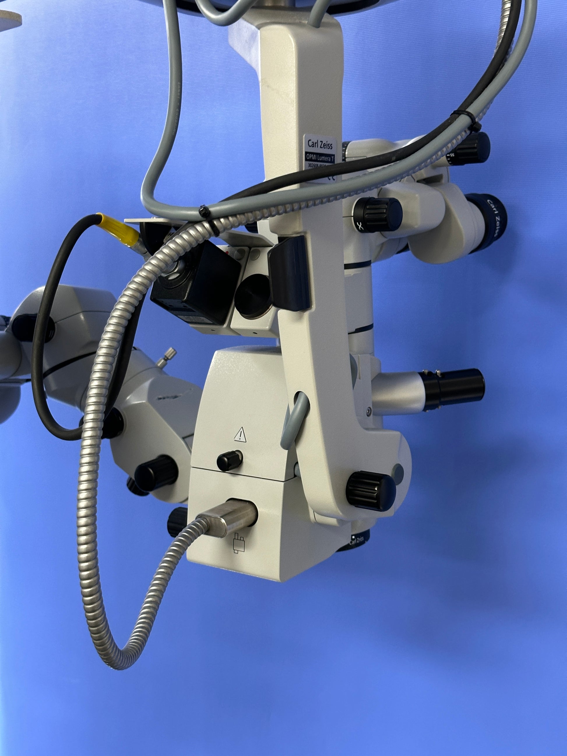

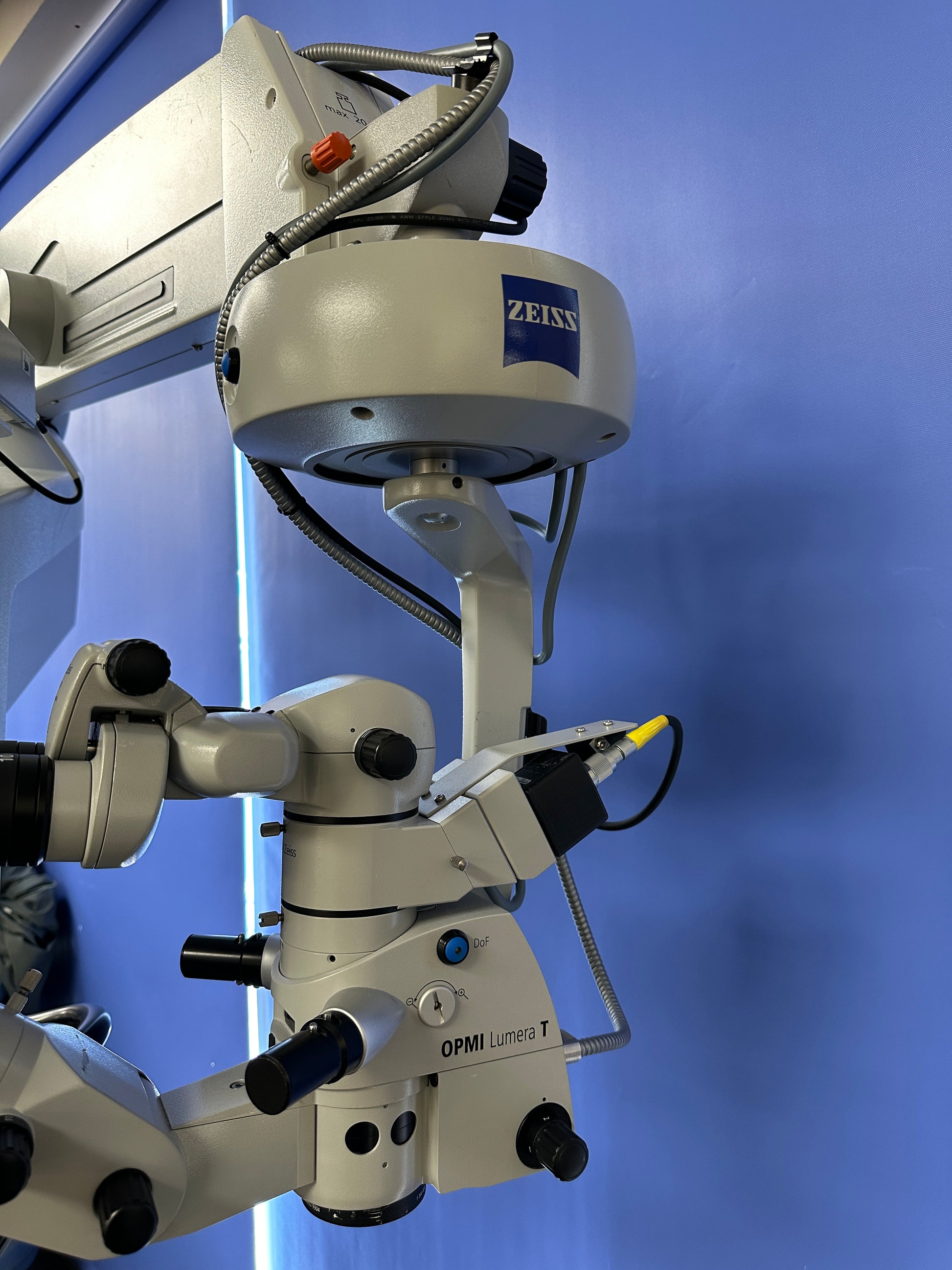

Zeiss OPMI Lumera T Dual Operated Surgical Microscope on S8 Base Unit
Zeiss OPMI Lumera T Dual Operated Surgical Microscope on S8 Base Unit
The Microscope includes the following
- 1 x Binocular
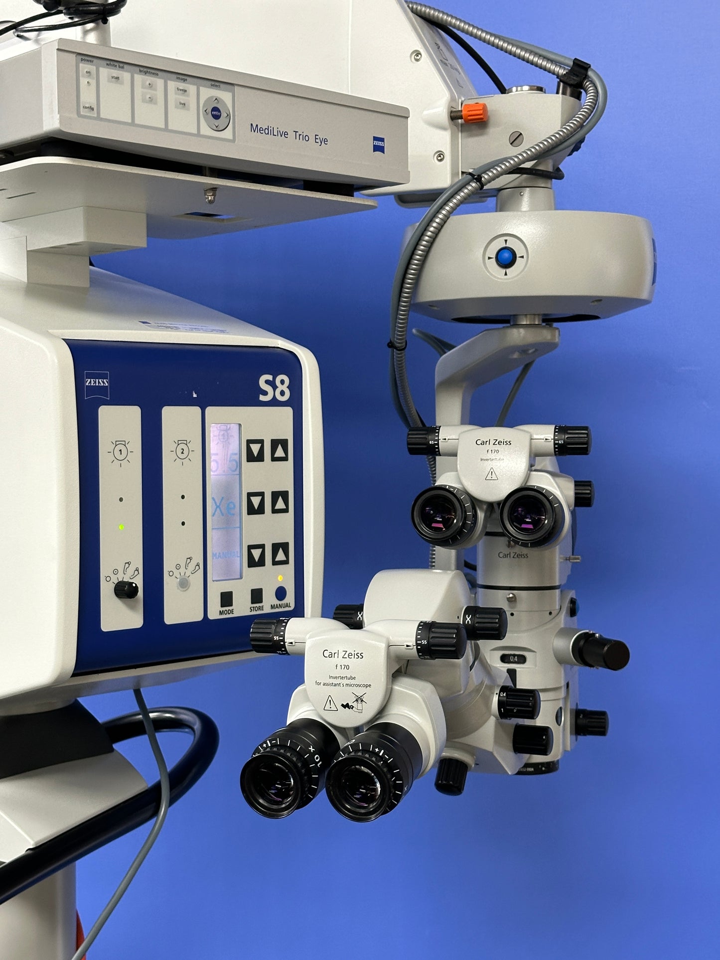
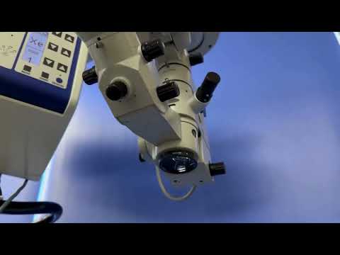
- Zeiss f170 Inverter Tube Microscope Head
- 2 x 10x Eyepieces
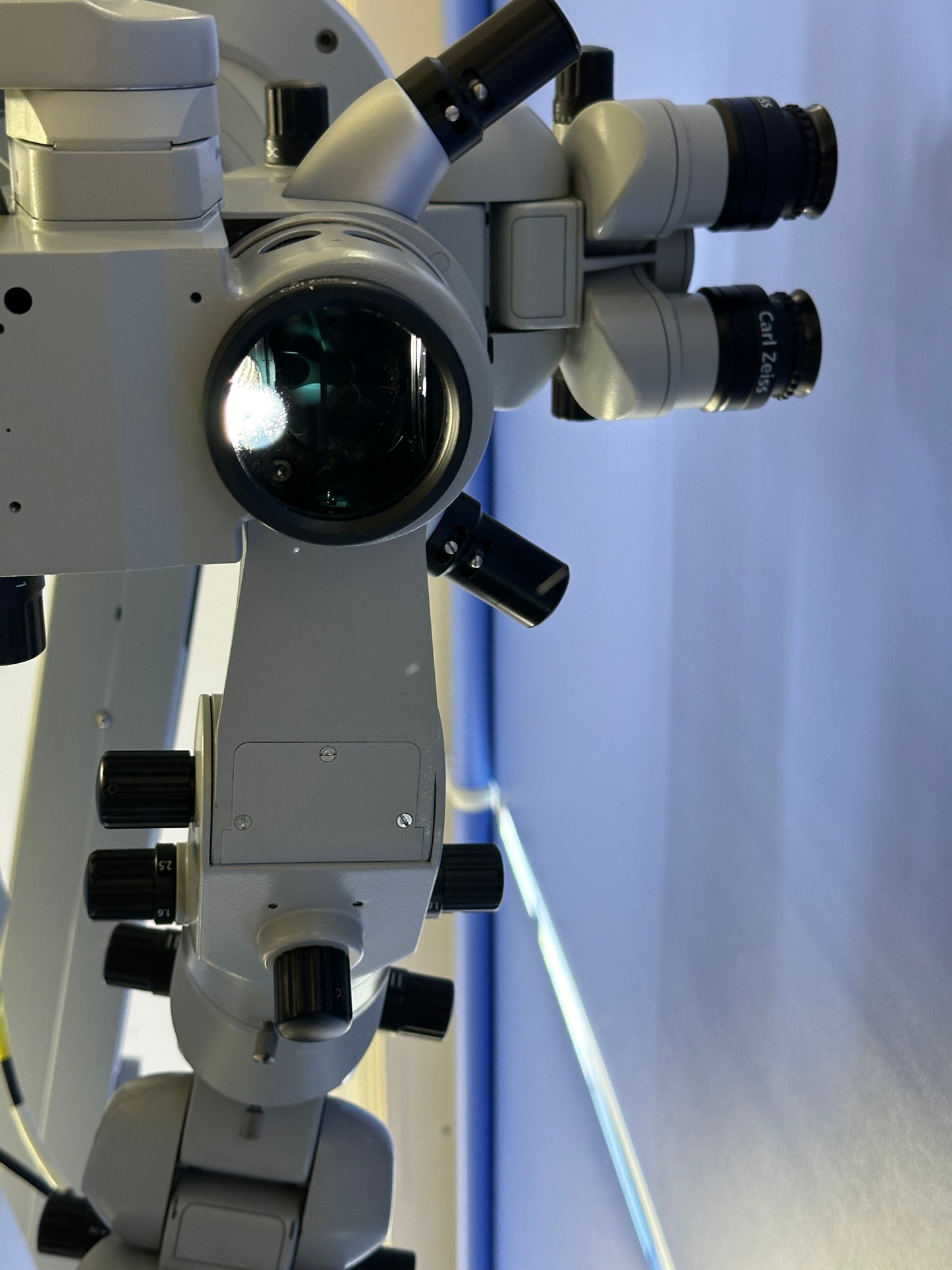
- Zeiss f 200 APO Lens
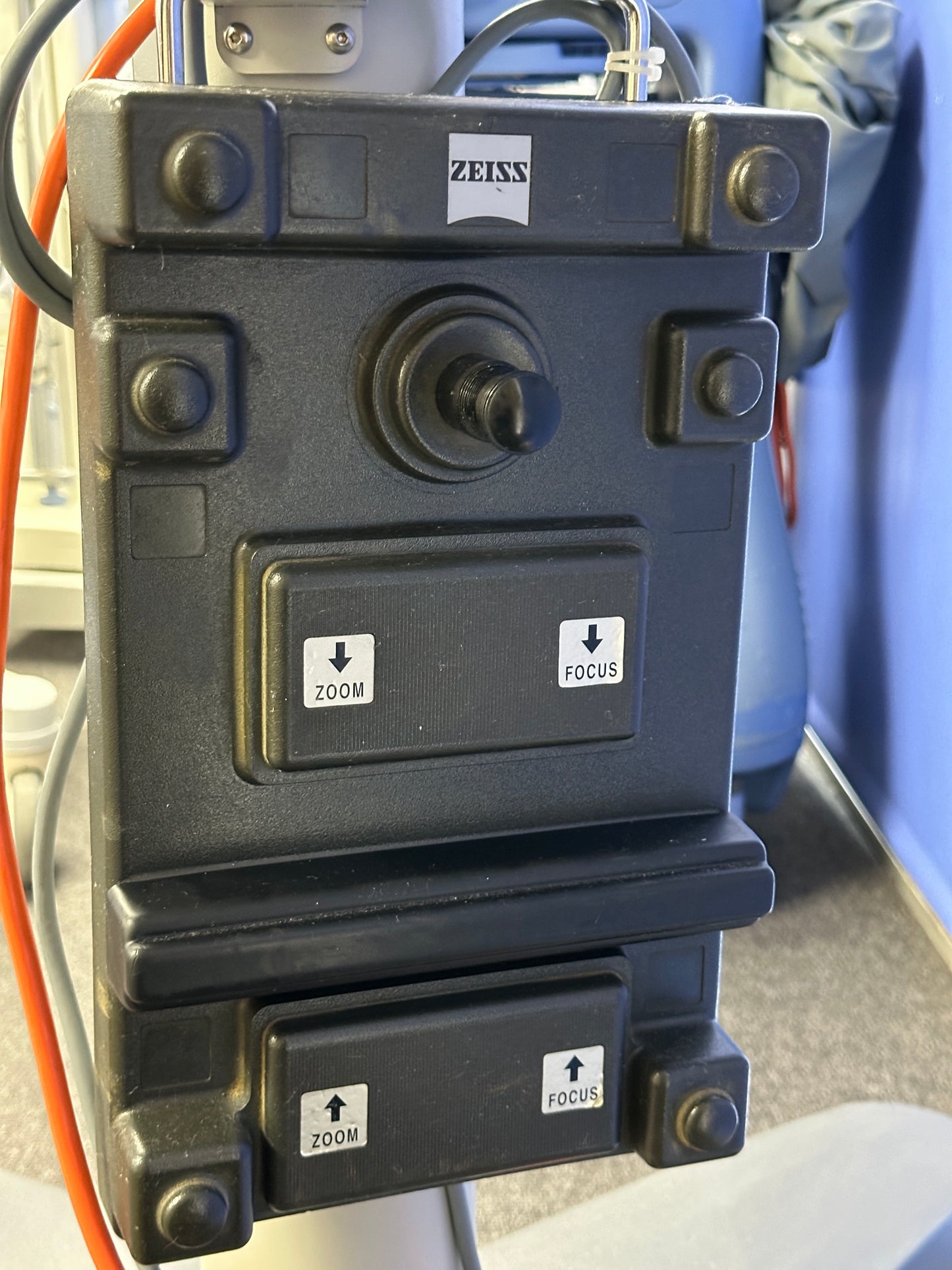
- Footswitch
- Zeiss MediLive Trio Eye Camera Control Unit
- Medicapture MediCap USB300 Medical
- Video Recorder
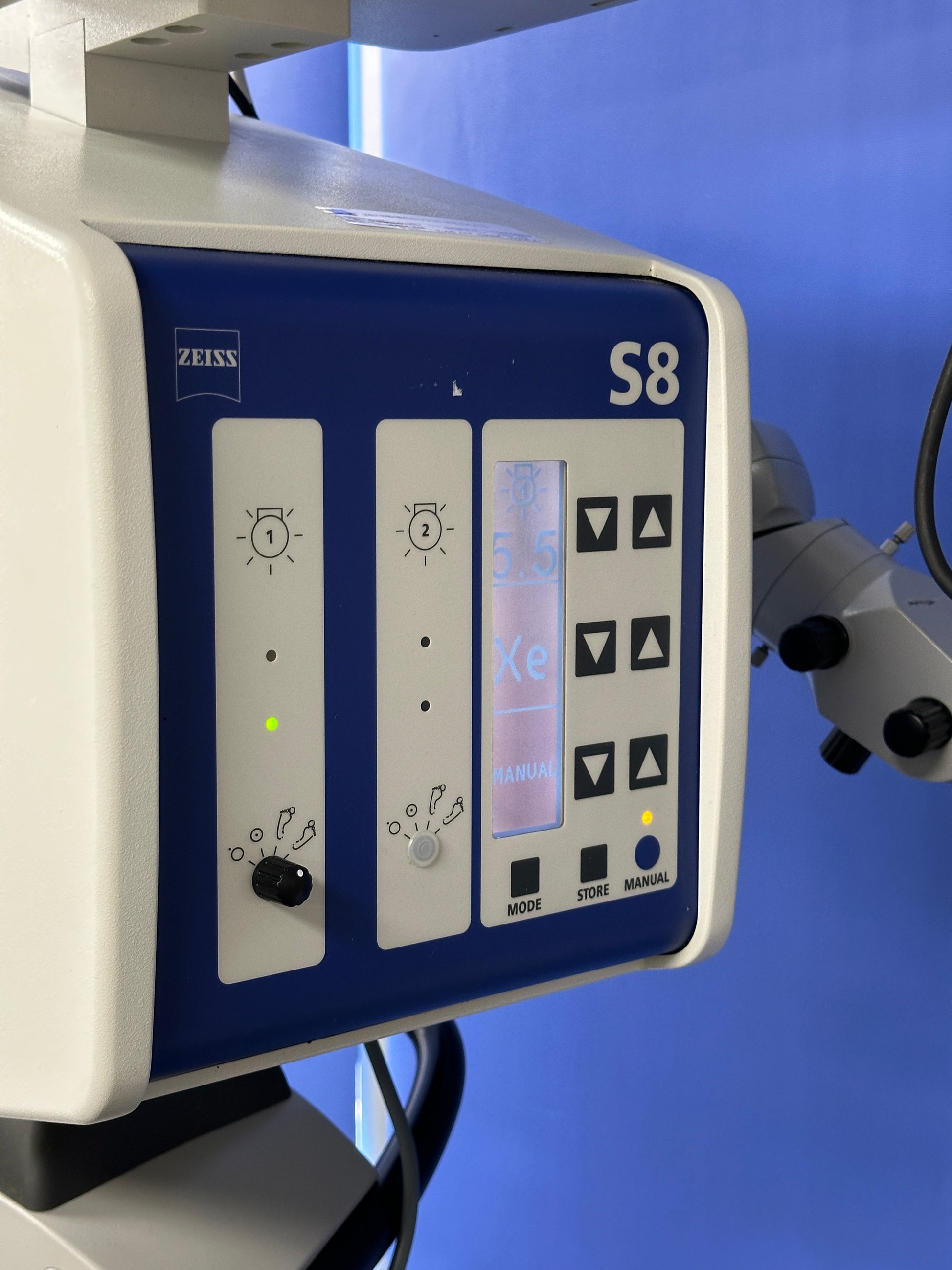
- Zeiss S8 Stand
Apochromatic: (three different frequencies to a common focus)
The apochromatic optics of the surgical microscope provide superb optical quality. The microscope image displays optimum contrast and excellent detail recognition along with a large depth of field.
The OPMI Lumera T surgical microscope can be used in all ophthalmic surgical procedures. The optional integrated assistant’s microscope makes it the ideal solution for teaching institutions.
The Zeiss Lumera T can be equipped with a completely integrated assistant’s microscope. Stereo Coaxial Illumination (SCI) reveals the fine details and effortless positioning with magnetic brakes provide handling comfort – quality made by Zeiss.
- See the most minute structures during surgery
- Identify details of the retina
- See overlays of the live image in the eyepiece with the External Data Injection System (EDIS)
- Manage depth of field with the push of a button
- View structures in the eye in natural colors
The 1Chip HD camera system:
with an integrated monitor for viewing videos provides excellent visualization of natural color renditions and crisp anatomical details.
Unmatched ZEISS optics:
for exceptional clarity, contrast and light.
RESIGHT from ZEISS
provides a clear, detailed view of the retina.
Instant Red Reflex :
brightly illuminates the eye – due to Stereo Coaxial Illumination (SCI) even with mature cataracts.
Integrated Superlux eye
xenon illumination allows you to view the structure of the eye in natural colors and high detail.
Integrated assistant microscope:
Focus and magnification are selected independently from the surgeon’s view, enabling active assistance.
Effortless positioning:
with magnetic brakes. The system smoothly glides into a new position. When locked, the surgical microscope remains firmly in place.
Deep View depth of field:
management system allows you to choose between maximum depth of field or optimum light transmission.
Assistance functions in the eyepiece:
Incision / LRI assistant:
Superimpose the exact position and size of the incisions to ensure precise 1,2,3 surgery.
Rhexis assistant:
Superimpose the exact shape and size of the capsulorhexis and center the IOL along the optical axis of the patient‘s eye.
Z ALIGN – toric assistant:
Inject reference axis and target axis in your microscope eyepiece to ensure precise 1,2,3 toric IOL alignment without corneal markers.
K TRACK:
Visualize corneal curvature in combination with a keratoscope,
e. g. in corneal transplantations.
Foot control freedom:
The foot control panel offers positioning flexibility and the ability to configure functions based on preferences.
External Video Components:
Video Cameras
TRIO 610 with CCU TRIO 600 – 3 Chip HD Camera System:
The high definition camera system with apochromatic video optics allows surgical microscope images to be generated with enhanced resolution and color rendition. The camera can be used for information, documentation, teaching and presentation of high quality images.
MediLive Trio Eye – 3 Chip SD Camera System:
Standard definition video camera with high sensitivity especially designed for different light conditions in anterior and posterior segment. The video images can be used for information, teaching, and documentation purposes. The MediLive Trio Eye helps the surgeon to easily overcome the challenges of ophthalmic imaging.
MEDIALINK 100:
Standard definition video recorder with image data management, that enables surgeons to record videos and capture still images. Using the MEDIALINK 100 standard definition videos and images can be automatically transferred to USB storage media or file servers.</p
HD Video Recorder:
A medical grade high definition video recorder is available.
HD Video Monitors:
Medical grade HD video monitors (1080p) are available in different sizes that match the surgical microscope video solutions.
Technical Data for Zeiss Lumera T Microscope:
· pochromatic Optics
· Motorized zoon system, 1:6 zoom ratio
· Focusing range: 50 mm
· Binocular tube: invertertube (optional 0-180° tiltable tube)
· Objective lens f = 200 mm (f = 175 mm optional)
· DeepView: depth of field management system
· Integrated fully stereoscopic assistant’s microscope with no light loss for the main surgeon
Illumination:
SCI: red reflex illumination and surrounding field illumination, both dimmable, patent pending
- Integrated 408 nm UV barrier filter
- Blue blocking filter
- Retinal protection device
- Fiber optic illumination
- Optional: fluorescence filter
Light source:
Superlux Eye xenon illumination with manual bulb change, including HaMode™ filter
Halogen illumination with fully automatic bulb change in case of lamp failure
Option: dual light source
Halogen + Halogen
Xenon + Halogen
X-Y coupling:
· 40 mm x 40 mm adjustment range
· Button for starting positions of the
· X-Y coupling and focus
Suspension System:
· S88 floor stand
· S8 ceiling mount
· S81 ceiling mount
The operating microscope powers up and initializes with LCD screen and keypads working
Both xenon lamps and brightness control are in good working order.
The optical head is intact with objective lens, binocular and eyepieces in place and the system produces clear images from both binoculars.
The suspension arm and electromagnetic brake release are working accordingly. Mechanical locks are also functional.
The footswitch operated motorized zoom, focus and X-Y movement are all working.
Z - mechanical adjustment is working as well. Centre positioning is working.
Fibre optic light guide cable is in good condition and transmission is good.
Camera system with MediLive Trio Eye and monitor display are included and working. Monitor display is in excellent cosmetic condition.
Our team of product experts ensures that each system is in excellent condition and produces stunning images when tested with a telescope.
Contact us today to learn more and get a personalized estimate for your needs.
Don't hesitate to ask any questions - we are here to help you make the most informed decision for your medical practice.
Share