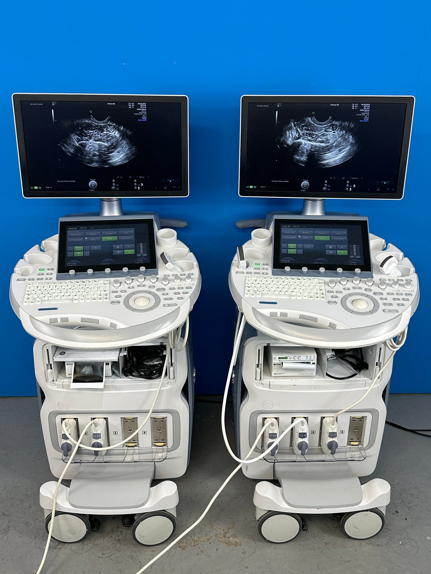
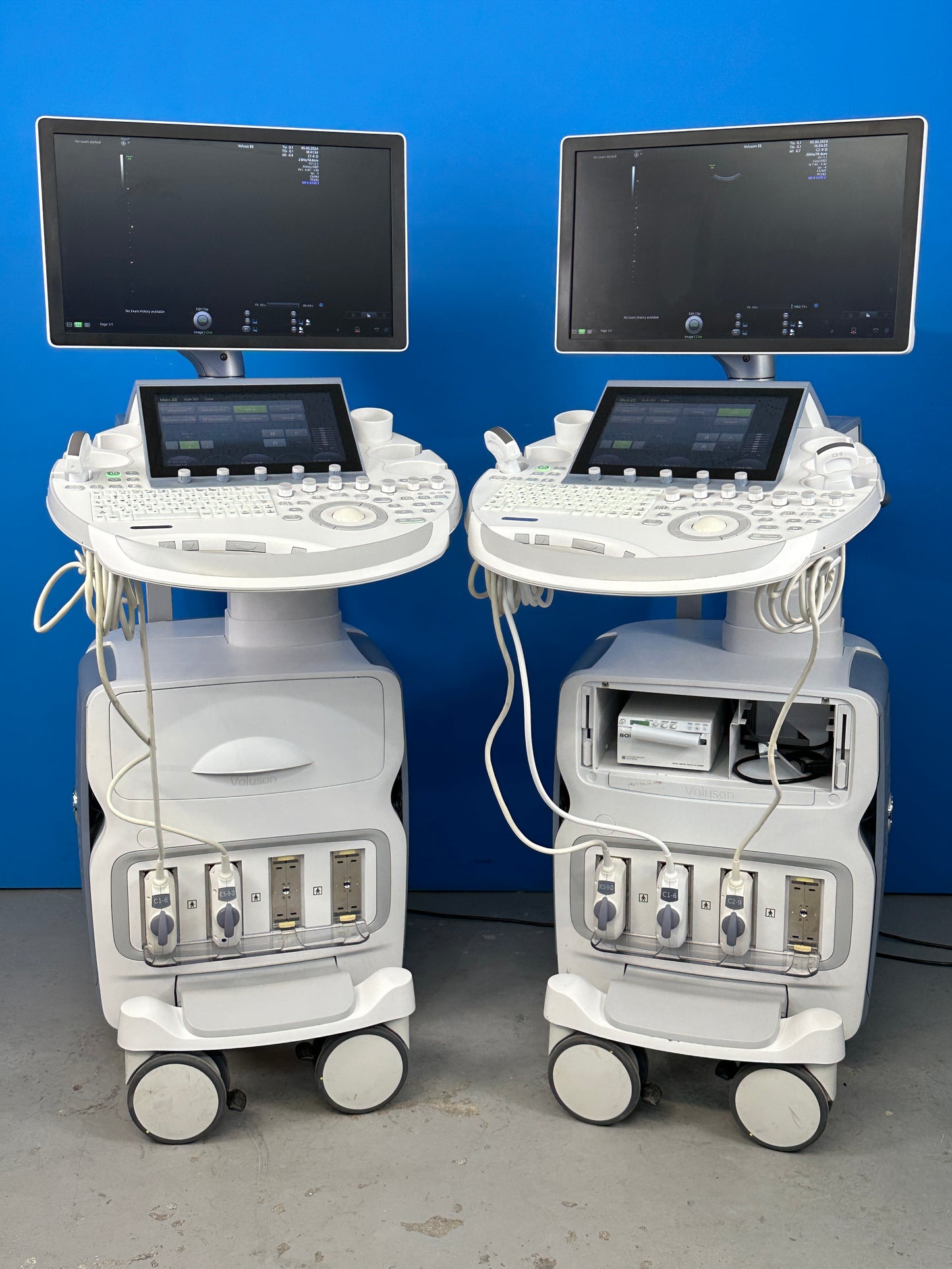
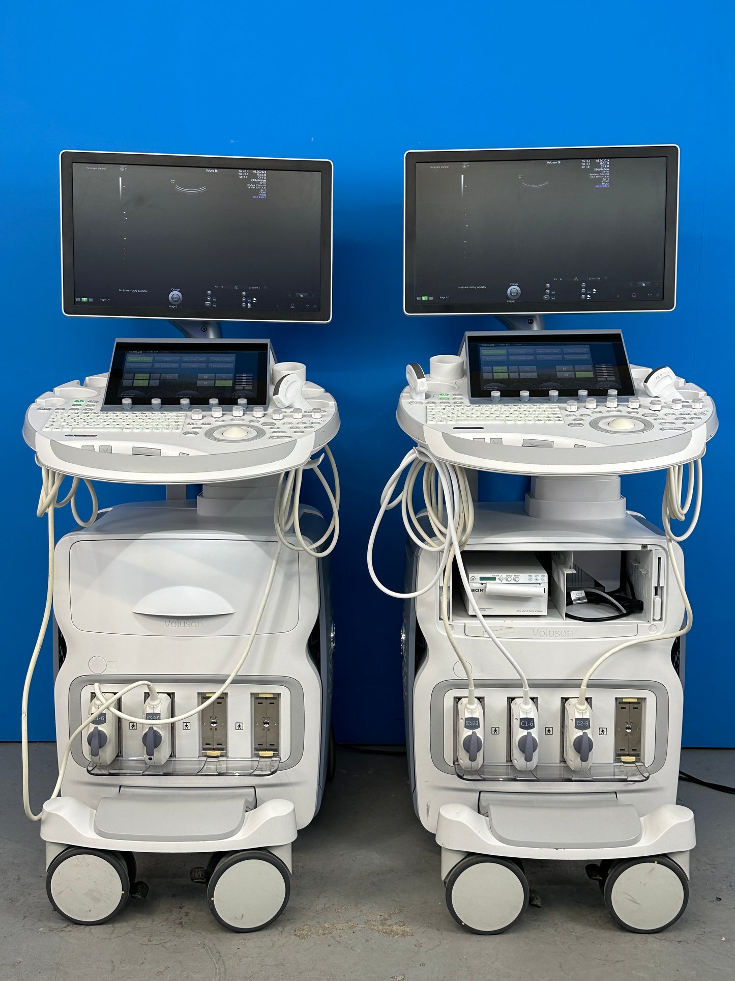
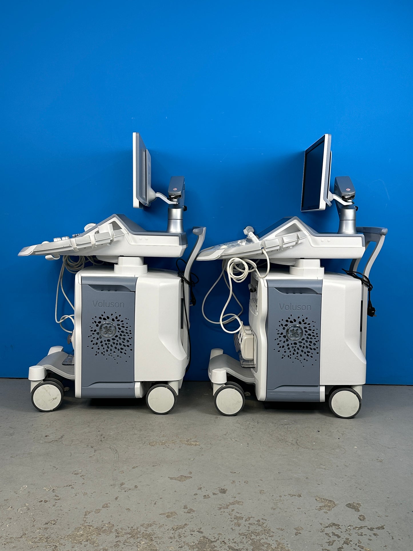
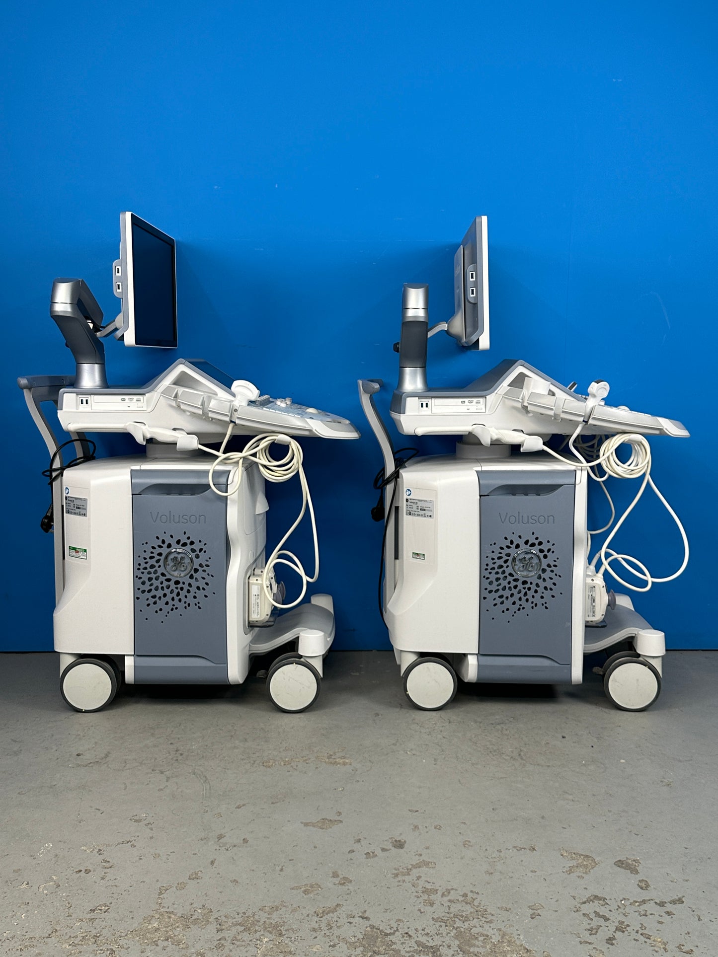
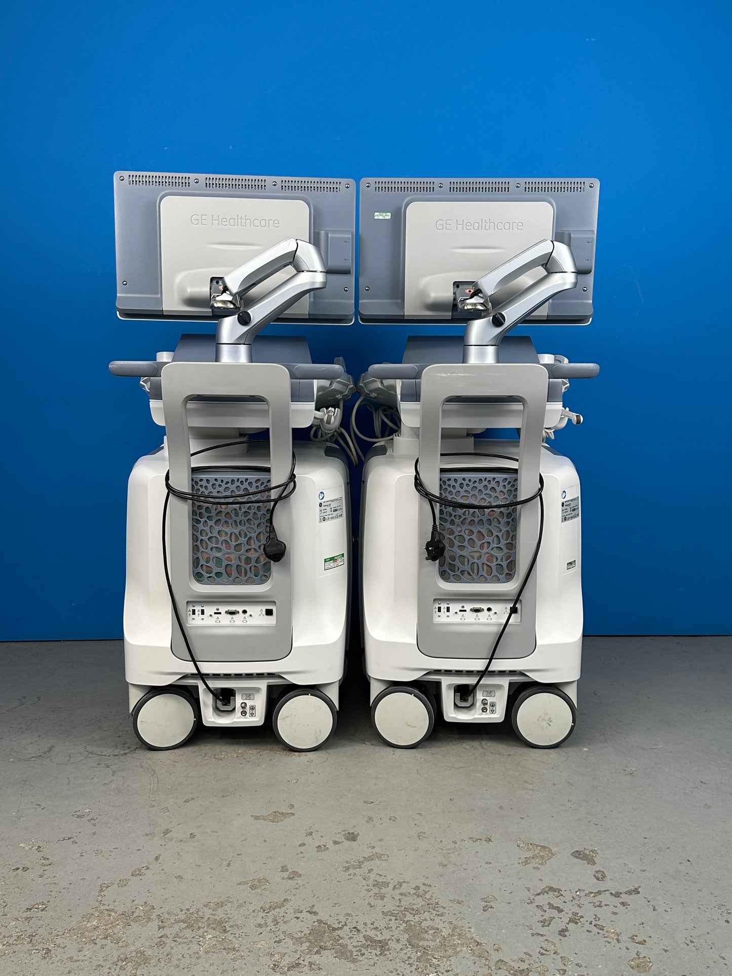
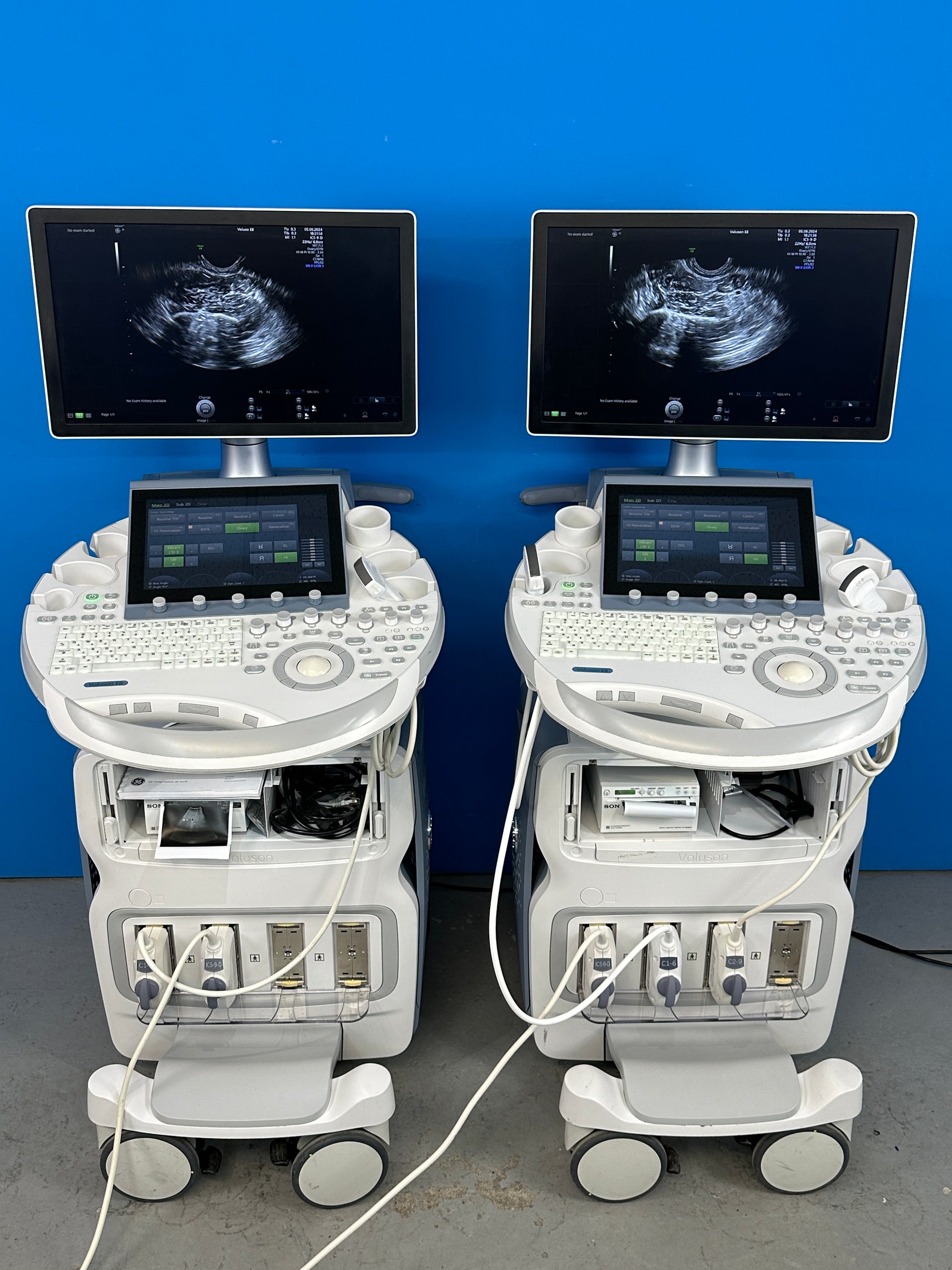
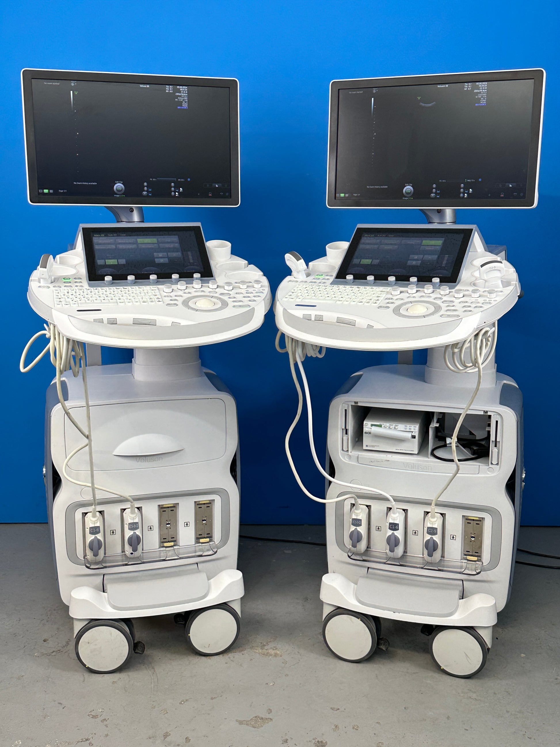
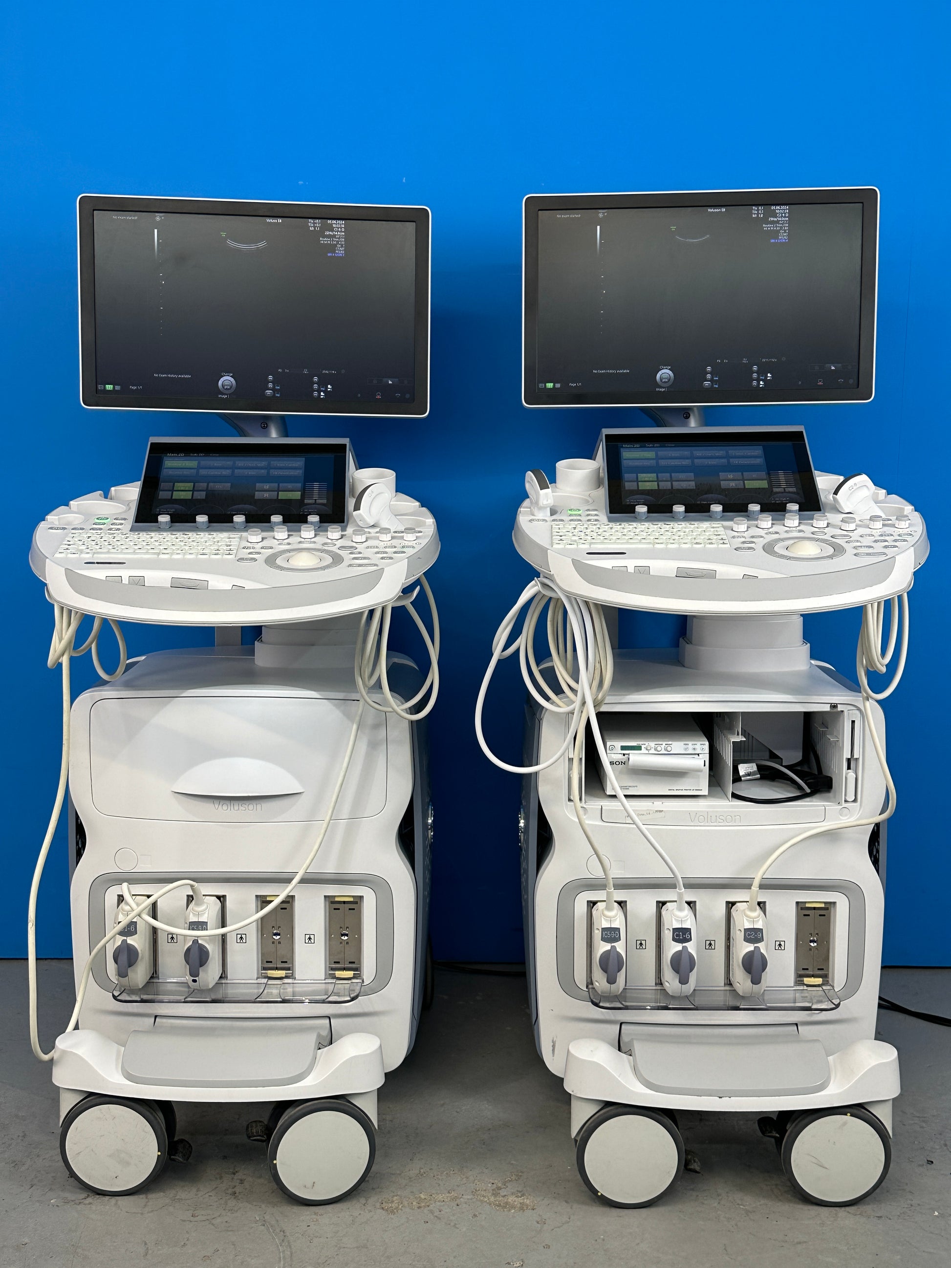
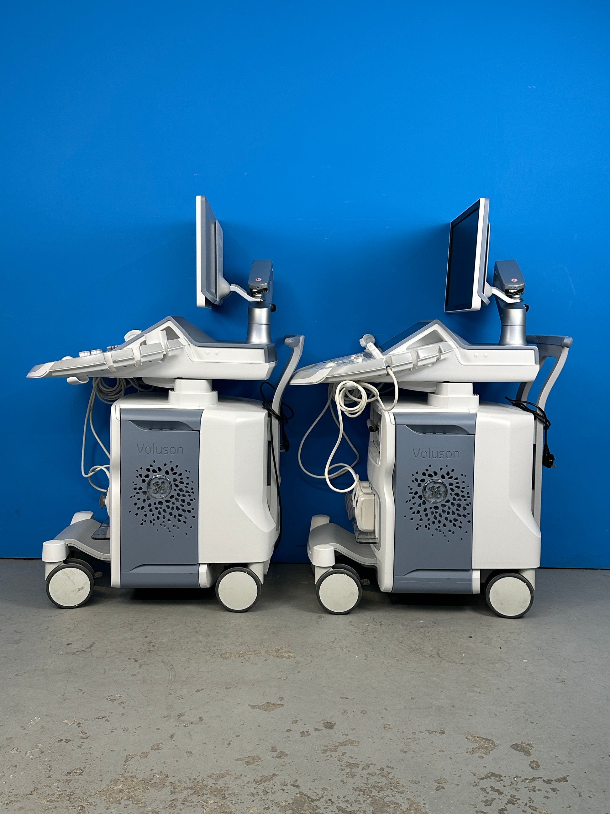
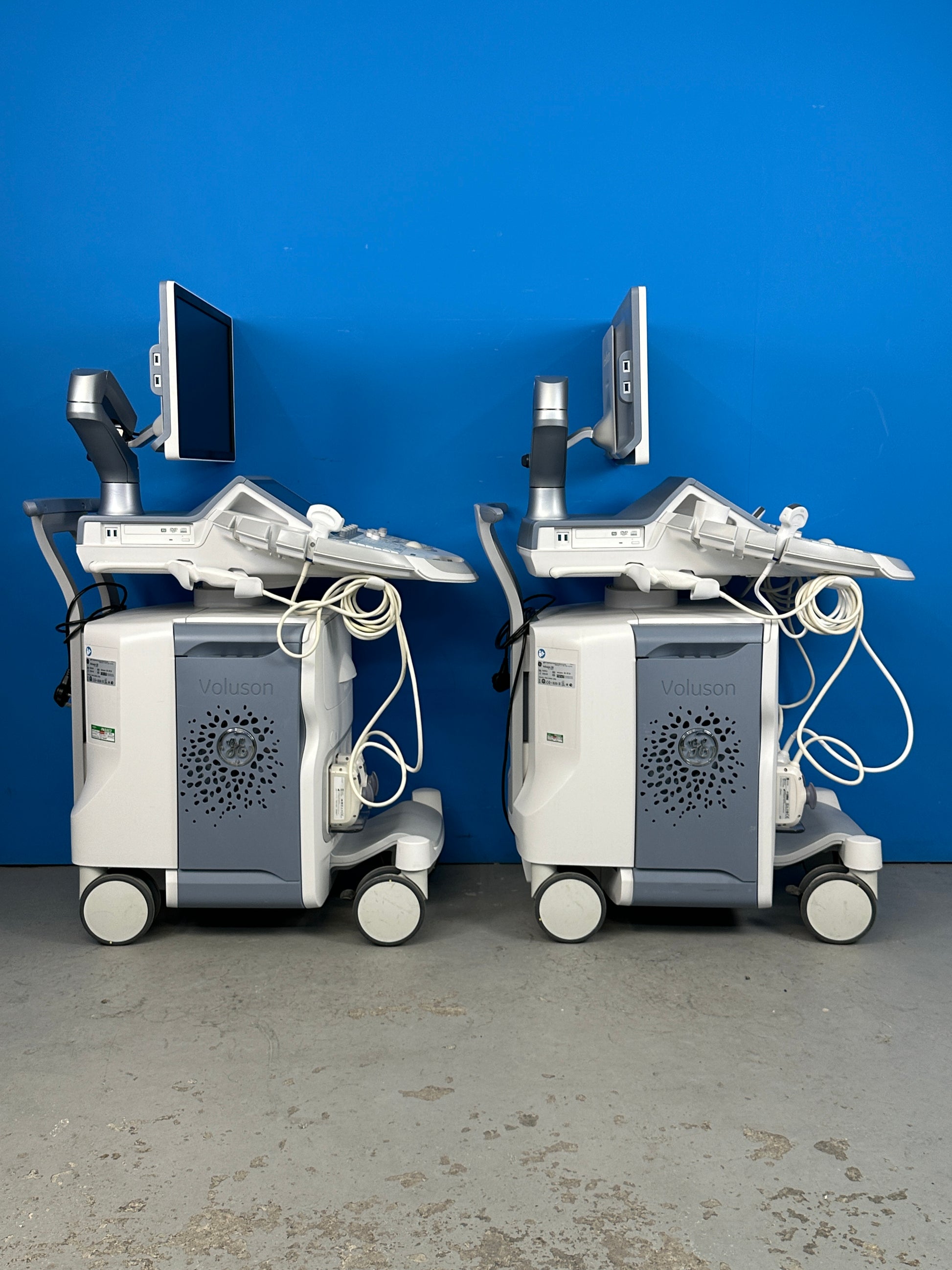
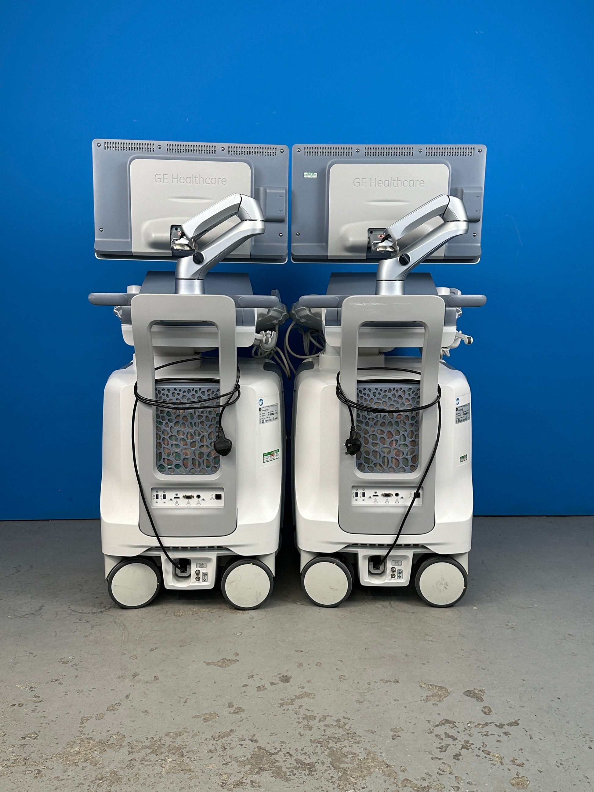
GE Voluson E8 BT19 Ultrasound System
GE Voluson E8 BT19 Ultrasound System
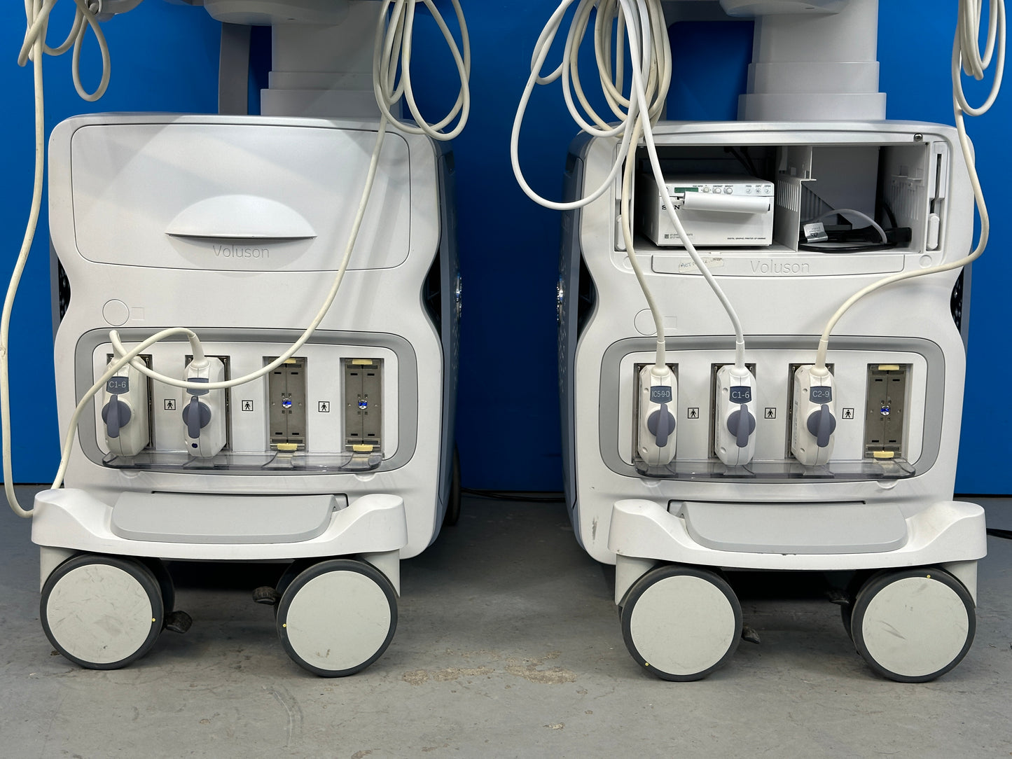
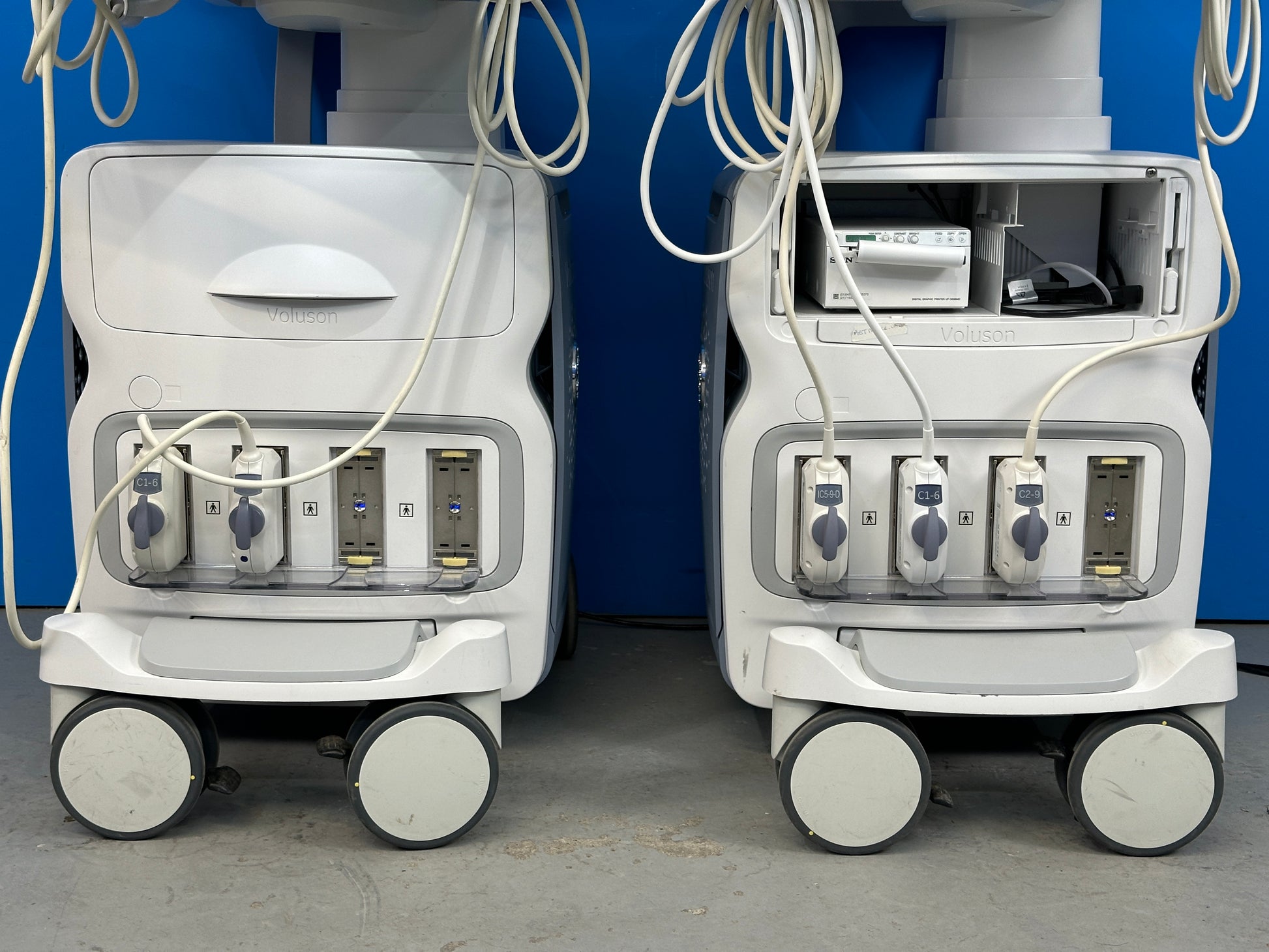
The ultrasound includes the following:
- C1-6-D XDClear Wide Band Convex Probe
- C2-9-D XDClear Wide Band Convex Probe
- Sony printer
The Voluson* E8 BT19 is an advanced imaging platform that combines extraordinary image quality with our superb volume ultrasound technology.
Features:
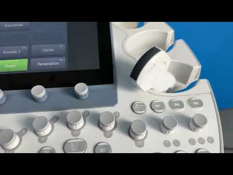
- Easy one-touch button workflow – Commonly-used scanning modes are right at your fingertips on the main screen of the touch panel
- Easy 3D and 4D imaging with automated tools and intuitive workflow
- Now you can adjust brightness with one touch for quick fine-tuning of image
- Enhance efficiency with 23'' widescreen LED monitor.
- Customizable layout to match your workflow needs, report preview enabling quick view on diagnostic measurements
- Image presentation in Standard or XL format allows you to see tiny details clearly
- One touch responsiveness. Simplified workflow.
- 1'' touch panel
- Auto TGC for quick fine-tuning of images
- Efficient menu navigation with swipe technology
- Quick and easy 1-button control panel up/down function allows for excellent ergonomics.
- Probe port illumination.
- Fast, secure data management for efficient communication.
- Integrated Software Digital Video Recorder, including
- USB recording
- Fast USB 3.0 connectivity
- Easy DICOM® integration
Exam applications:
- Abdominal
- Obstetrical Gynecological
- Small Parts and Breast
- Vascular
- Pediatrics
- Transrectal
- Cardiology
- Cephalic
- Musculoskeletal (MSK)
Operating modes:
- Brightness Mode (B-Mode) (2D)
- Motion Mode – M-Mode (conventional M-Mode)
- Anatomical M-Mode (AMM)
- Pulsed Wave Doppler (PW) with HPRF
- Continuous Wave Doppler imaging (CW)
- Color Flow Doppler mode (CFM)
- Power Doppler Mode (PD)
- High Definition Power Doppler (HD-Flow*)
- Tissue Doppler Mode (TD) and PW-Tissue Doppler Mode
- B-Flow (BF)
- Compression & Shearwave Elastography (May not be available in all countries)
Contrast Imaging Mode Combination modes:
M/CFM, M/HD-Flow, M/TD, PW/CFM, PW/HD-Flow, PW/PD, PW/TD Extended View (XTD View) Volume Mode (3D/4D):
- 3D Static
- 4D Real Time
- VCI-A
- VCI-OmniView
- Spatio-Temporal Image Correlation (STIC)
- 4D
HDlive:
This innovative rendering technology helps provide exceptional anatomical realism with increased depth perception to help enhance clinical confidence. HDlive has brought anatomical realism to the next level with the introduction of two new.
optional technologies:
HDlive Silhouette:
variable transparency reveals internal structures, helping you make a confident volume assessment of 1st trimester anatomy.
HDlive Flow:
Presents vascular structures with greater depth perception and anatomical realism
Advanced Volume Contrast Imaging (VCI) with OmniView
- Helps improve contrast resolution and visualization of the rendered anatomy with clarity in any image plane, even when viewing irregularly shaped structures
SonoRenderlive
- Helps enhance efficiency in volume rendering with automated placement of the render line for optimized surface rendering. SonoRenderlive continuously updates render line placement with fetal movement during 4D examinations.
The Ultrasound is in Excellent Condition.
Our team of product experts ensures that each system is in excellent condition and produces stunning images when tested with a Probe.
Contact us today to learn more and get a personalized estimate for your needs.
We are here to help you make the most informed decision for your medical equipment purchases in your budget.
Share
