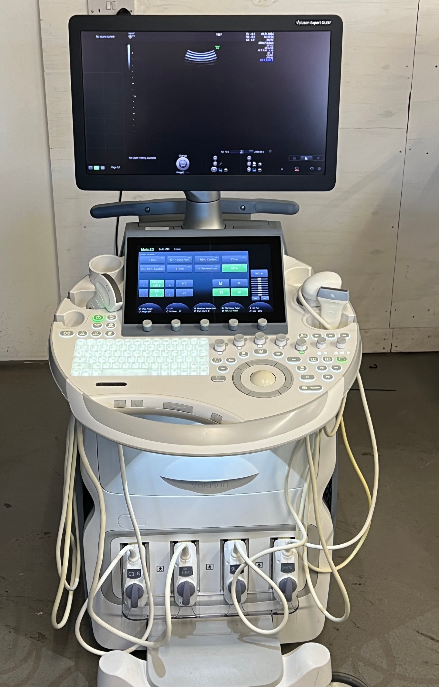

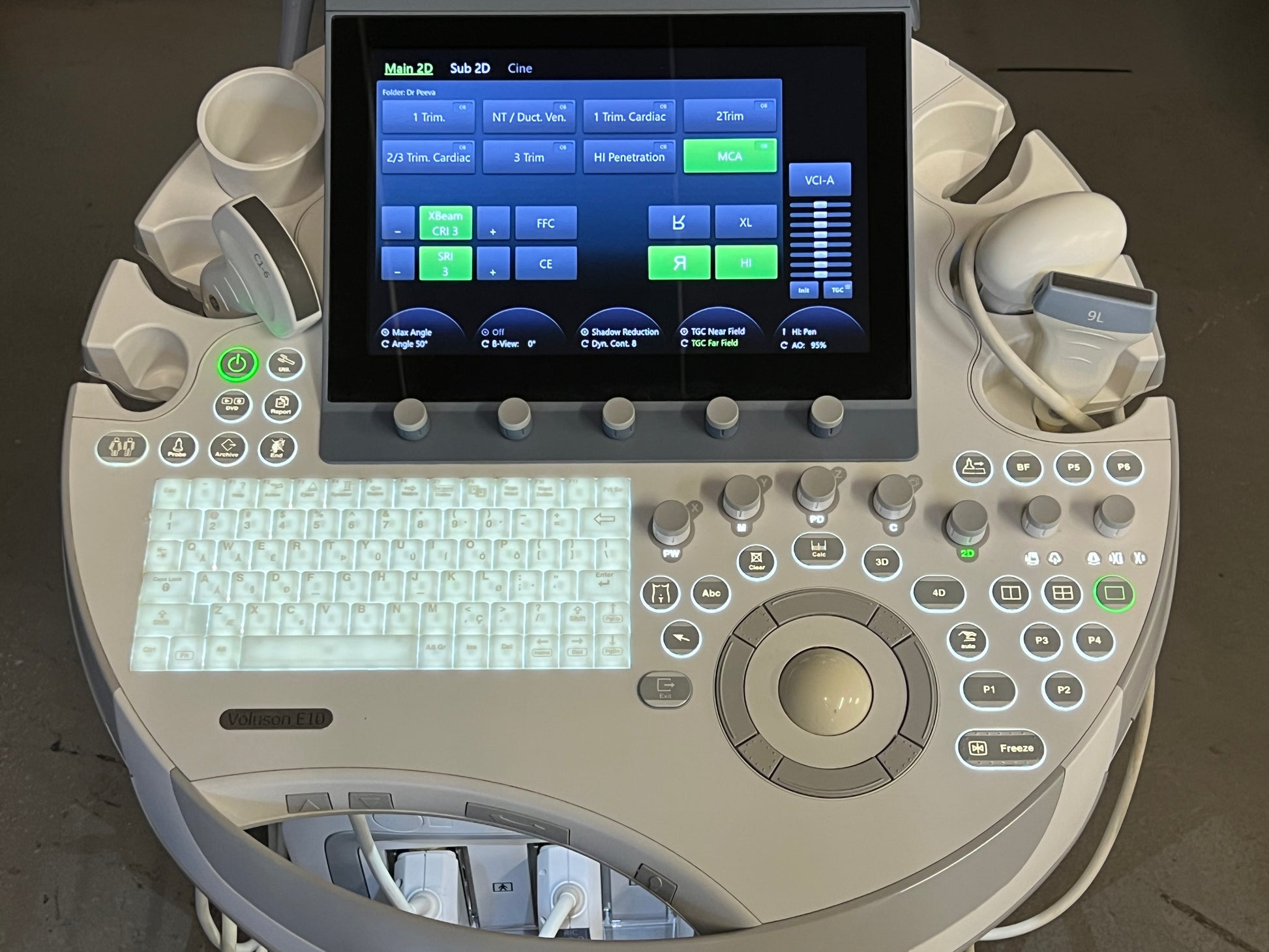


GE Voluson E10 Ultrasound with BT21
GE Voluson E10 Ultrasound with BT21
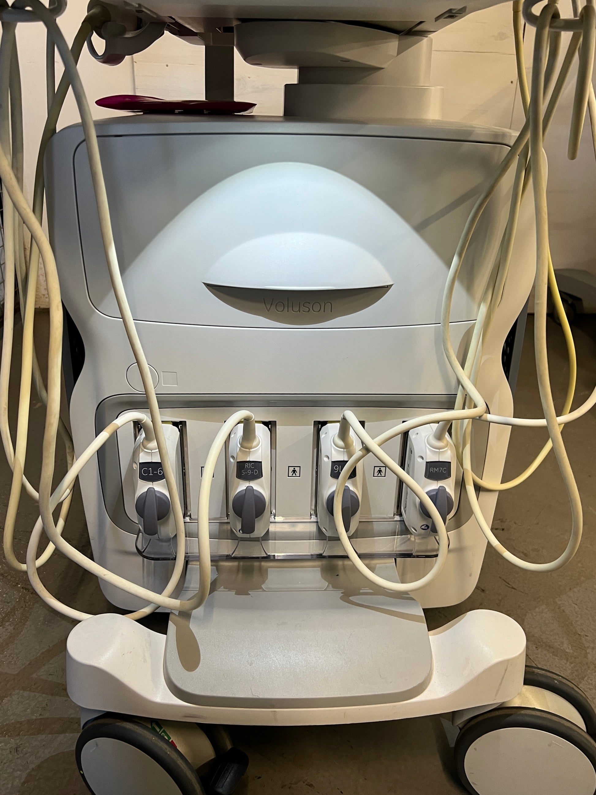
The Ultrasound includes:
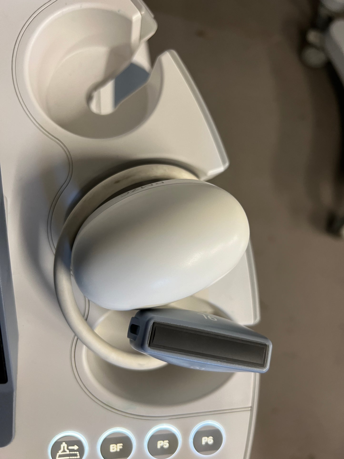
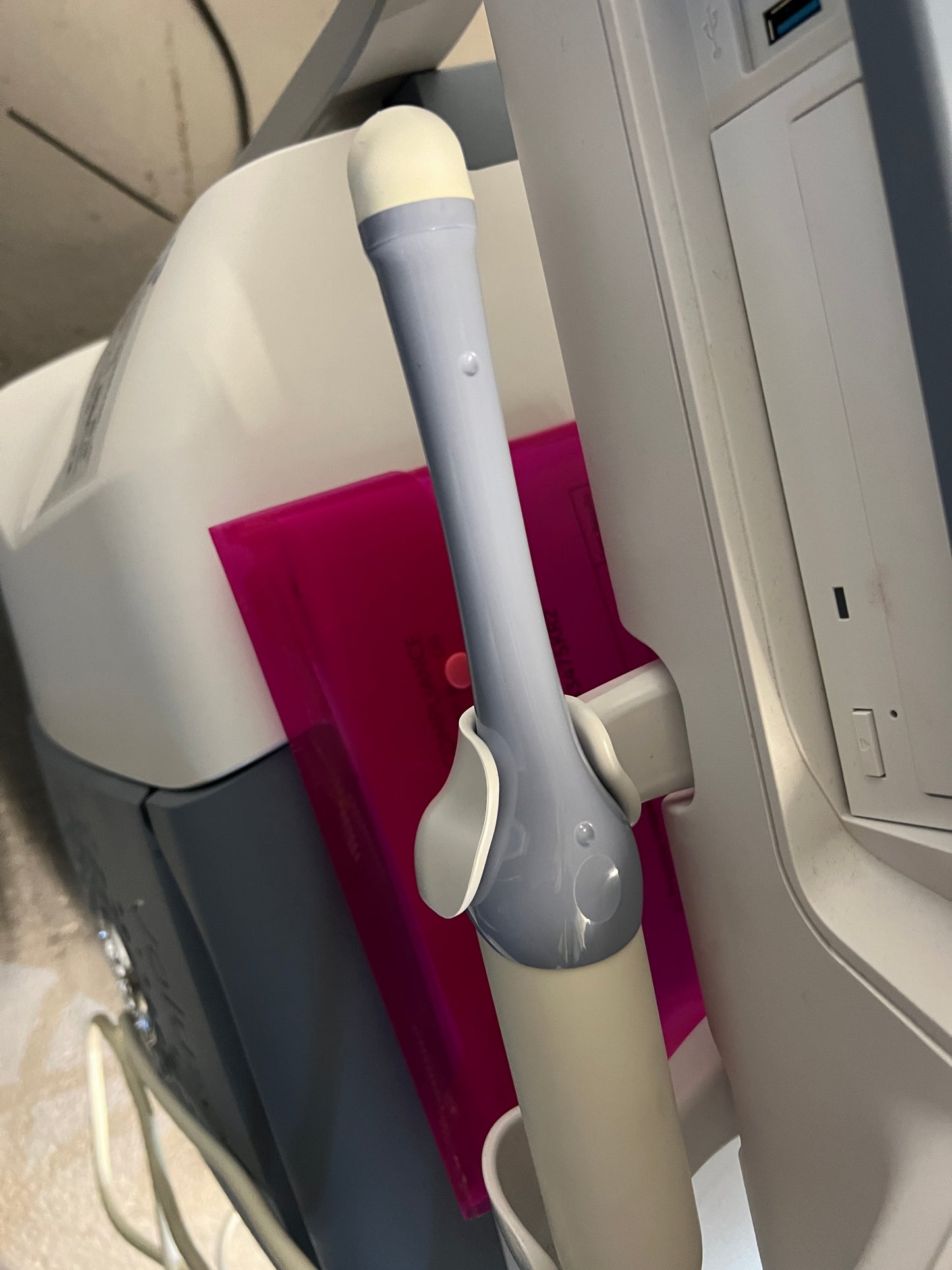
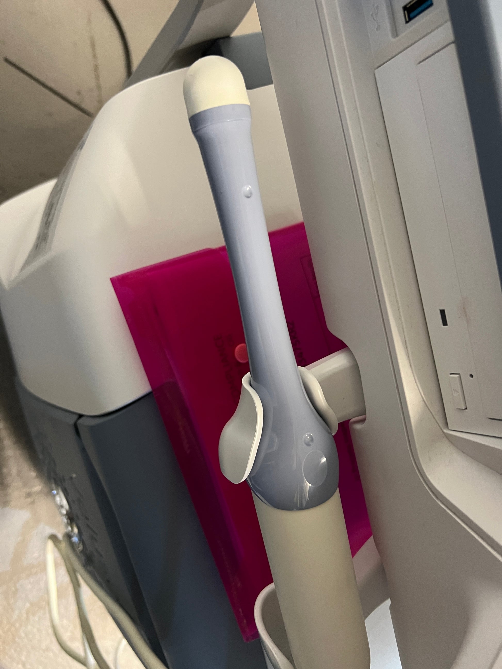
- Convex Probe
- Convex 4D Probe
- TVS 4D Probe
- Linear Probe
The GE Voluson E10 is an ultrasound designed for women’s health. The Voluson E10 is dedicated to providing ultrasonic images for obstetrics and gynecology. The Voluson E10 ultrasound machine can provide premium imaging with the Matrix 4D transducer. Four images are simultaneously taken at a higher frame rate to create a more accurate 4D image. The Voluson E10 ultrasound’s operating system runs a Windows 10 platform and has a 1TB hard drive for storage, along with USB ports for storage output. With a 23-inch monitor to view the crisp, realistic live images. Along with a 12-inch touchscreen control panel. The Voluson E10 ultrasound machine can additionally support abdominal, pediatric, small organ, cephalic, and peripheral vascular imaging.
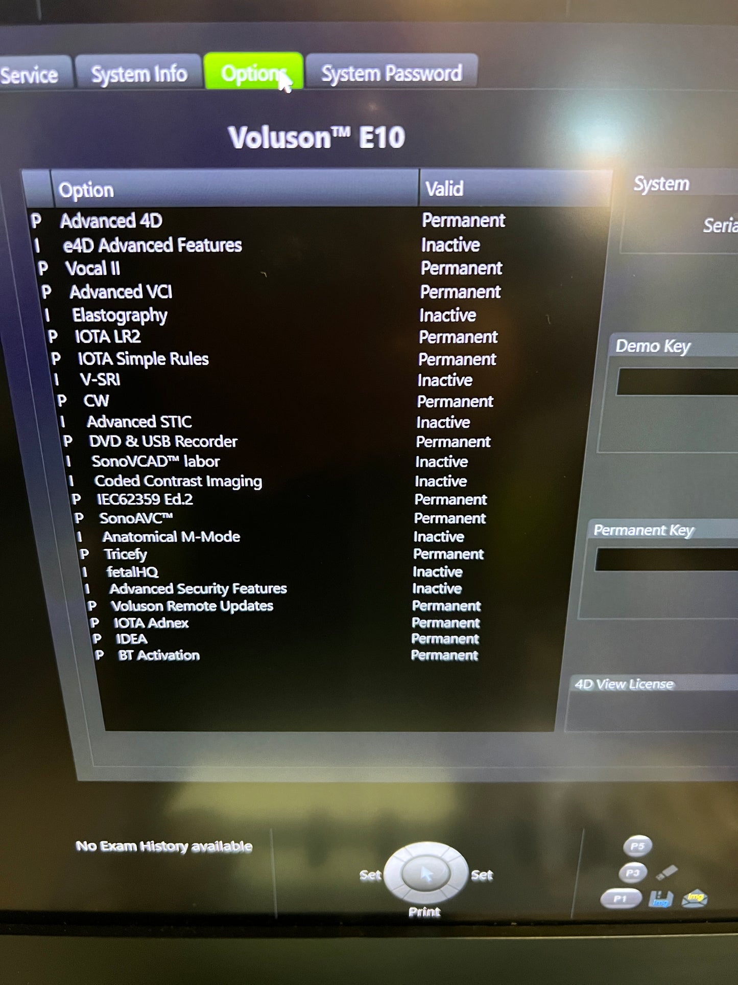
Key Features:
- Biplane imaging – Display high resolution and high frame rate images in two perpendicular planes for exceptional quality.
- VCI-A (Volume Contrast Imaging) – Creates high contrast resolution with grayscale and color Doppler images
- eSTIC (Spatio-temporal image correlation) – Produces up to a 75% reduction in acquisition time over traditional STIC for fetal cardiac exams
- e4D SnapShot – Real-time 4D for higher resolution 3D volume or eSTIC data sets
- Tricefy Inside capabilities
- Display more image detail and clarity in less time with electronic matrix 4D probe technology through ultra-fast frame rates, flexible imaging formats, and excellent resolution
- Boost exam efficiency and accuracy through Voluson automation tools
MORE CLARITY:
Ultrasound Pathways for spectacular 2D and 3D/4D images with increased penetration. MORE SPEED the data transfer rate for higher resolution and very fast frame rates. MORE FLEXIBILITY the processing power for advanced applications and efficient workflow. BT21 brings very strong and relevant innovation – RM7C along with other IQ tunings, such as introduction of Shadow Reduction, emphasize the focus on enhancing imaging performance. In Gynecology, additional auto-sequence measurements and Scan Assistant protocols simplify the valuation of the pelvic floor.
Electronic 4D:
Radiance System Architecture, along with the eM6C probe delivers ultra-fast volume rates, flexible imaging formats and excellent resolution from routine OB exams to complex fetal echocardiography.
- Bi-Plane
imaging provides simultaneous display of high resolution, high frame rate images in two perpendicular plans
- VCI-A
(Volume Contrast Imaging) delivers excellent contrast resolution through thick slide volume of gray scale and color Doppler images
- eSTIC
(Spatio-Temporal Image Correction) enhances fetal cardiac exams with up to 75% reduction in acquisition time over traditional STIC
- e4D
SnapShot optimizes you exam time with one button access from Real-time 4D to acquire high resolution 3D volume or eSTIC dataset.
V-SRI
Improve 3D/4D quality in multi-planar studies to enhance smoothing effect on rendered images through speckle reduction.
Advanced VCI
Adjusts slice thickness on 3D or 4D images to help enhance contrast resolution with use of render techniques such as bone and tissue renderings. Can be applied in the acquisition plane (VCI-A), static 3D volumes or OmniView.
OmniView
Obtain any plane from a 3D or 4D volume by simply drawing a line, curve, poly-line or trace through a structure. This valuable technology enables views of even irregularly shaped structures not attainable in 2D imaging.
SonoRenderlive:
Facilitate volume rendering with an automated placement of the render line for optimal surface rendering. SonoRenderlive continuously updates render line placement with fetal movement during 4D examinations
HDlive Technologi:
HDlive Silhouette:
Dynamically apply targeted transparency to rendered structures for a more comprehensive view of anatomy from solid surface structure to developing internal anatomy
HDlive Studio:
Illuminate anatomy and fluid with up to three independent light sources of variable color, intensity and direction to focus on even the tiniest of structures.
HDlive Flow:
Clearly display vascular structures with greater dimension – from small vessels to the great arteries.
HDlive Flow Silhouette:
Visualize blood vessels from a surface or targeted transparent view to provide greater insight into vascular anatomy and surround structures.
Application:
A remote control for the heater-cooler unit permits patient temperature control and monitoring without diverting the perfusionist's attention.
- Breast
- General Imaging
- OB/GYN
- Vascular
The Ultrasound is in Excellent Condition.
Our team of product experts ensures that each system is in excellent condition and produces stunning images when tested with a telescope.
Contact us today to learn more and get a personalized estimate for your needs.
We are here to help you make the most informed decision for your medical equipment purchases in your budget.
Share